20.12 – Phylogenetically independent contrasts

Introduction

Assumption of independence among the subjects in a study, key assumption. Cross species comparisons as tests of correlated evolution of traits has a long history in biology research (Cornwallis and Griffin 2024). Methods have transitioned from observations to rigorous, phylogenetic tree-based statistical approaches (phylogenetic comparative methods), and continue to provide important analytical tools, eg., host adaptation and cross-species transmission of Covid-19 virus (Attwood et al 2022).

Comparisons among species for the association between body size and metabolic rate, for example Kleiber’s Law, are classic cases (Fig 1, after Table 2 in Kleiber 1947). We’ll use Kleiber’s data basal metabolic rate (BMR) and body mass for a collection of species to explore two phylogenetic comparative methods, PIC and PGLS.
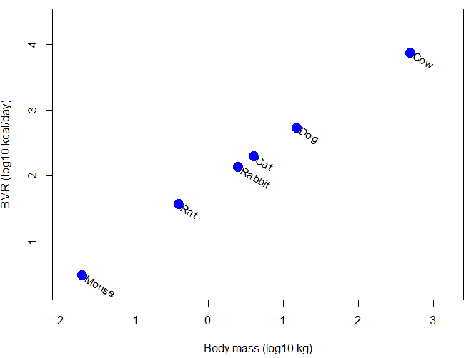
Scatter plot of BMR on Body Mass, log-transformed.

Figure 1. Scatter plot of BMR on Body Mass, log-transformed.

Typical approaches statistically, use of ANOVA or linear regression approaches. A basic assumption of ANOVA is that sampling units are independent 13.1 – ANOVA assumptions.

Prior to the 1980s, it was rarely appreciated in comparative analysis that species are not independent sample units (Harvey and Pagel 1991); evolution produced nested hierarchical relationships among the species. We recognize this with phylogenies (Felsenstein 1985, Harvey and Pagel 1991, Martins 1996, Garland et al 2005).  Mice and rats share more recent common ancestor, and cattle and pigs share more recent common ancestor, than do mice and cattle, for example. Felsenstein (1985, 1988), largely credited for making the argument that Type I error likely if phylogeny ignored, and, importantly, provided an algorithm, Phylogenetic Independent Contrasts, PIC, which provided a simple way to correct for phylogenetic nonindependence. Felsentein’s landmark 1985 paper has been cited more than ten thousand times (Feb 2024).

Logic of PIC

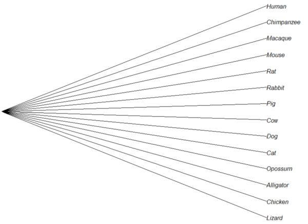
Treating comparative data, eg, species, as a collection of independent samples implies that the evolutionary history was a spontaneous burst, or star-like phylogeny — an unresolved phylogenetic tree with branches radiating from a single ancestral node (Fig 2).
star phylogeny

Figure 2. Star tree phylogeny (same image shown Figure 5, 20.11 – Plot a Newick tree).

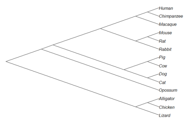
But what nature provides is nonindependence (Fig 3, for more about star phylogeny in PIC see discussion in Garland et al 2005), Darwin’s “groups under groups,” or a nested hiearchy. Species are not independent units and phylogenetic history should be accounted for during statistical analysis.
diagonal cladogram

Figure 3. A cladogram for same species, showing the hierarchical, nested relationships among taxa, what nature actually provides (same image shown Figure 2, 20.11 – Plot a Newick tree).

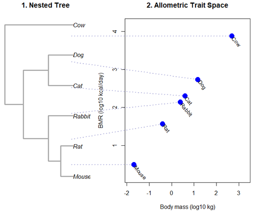
Let’s revisit Figure 1. What do the points look like if we line up the known phylogeny for Kleiber’s six species against the plotted data? That gives us Figure 4.
Kleiber's Law and the confounding effect of shared ancestry.

Figure 4. Kleiber’s Law and the confounding effect of shared ancestry.

Dashed connecting lines in Figure 4 trace each species from its tree position to its phenotypic value, demonstrating how phylogenetic clustering violates the independent data assumptions of standard OLS regression.

Begin R code for Figure 4, with remarks — code generated with Gemini AI assistance.

# 1. Reset the data frame 
kleiber_data <- data.frame(
Species = c("Mouse", "Rat", "Rabbit", "Cat", "Dog", "Cow"),
Mass = c(0.021, 0.282, 2.45, 3.1, 13.4, 439.0),
BMR = c(3.38, 32.4, 134.0, 151.0, 506.0, 6135.0),
stringsAsFactors = FALSE
)
kleiber_data$log_Mass <- log10(kleiber_data$Mass)
kleiber_data$log_BMR <- log10(kleiber_data$BMR)

# 2. Build the exact matching standalone 6-species tree
library(ape)
newick_tree_6 <- "((((Mouse:10,Rat:10):10,Rabbit:20):20,(Cat:20,Dog:20):20):15,Cow:55);"
my_tree <- read.tree(text = newick_tree_6)

# 3. Setup a side-by-side 2-panel layout
layout(matrix(c(1, 2), nrow = 1), widths = c(1, 1.3))
par(mar = c(5, 4, 4, 1))

# PANEL 1: Plot the tree hierarchy 
plot(my_tree, main = "1. Nested Tree", 
edge.width = 3, edge.color = "darkgrey", cex = 1.1)

# FIXED COORDINATE MAP: 'ape' plots leaves bottom-to-top exactly matching the tree order
# We map the tree tip labels to simple integers 1 through 6
tree_heights <- 1:6
names(tree_heights) <- my_tree$tip.label

# PANEL 2: Plot the scatterplot
plot(kleiber_data$log_BMR ~ kleiber_data$log_Mass, 
pch = 19, cex = 2, col = "blue", 
xlab = "Body mass (log10 kg)", ylab = "BMR (log10 kcal/day)",
xlim = c(min(kleiber_data$log_Mass) - 0.2, max(kleiber_data$log_Mass) + 0.6),
ylim = c(min(kleiber_data$log_BMR) - 0.3, max(kleiber_data$log_BMR) + 0.3),
main = "2. Allometric Trait Space")

# Re-apply your custom 310-degree slanted text labels
text(kleiber_data$log_Mass + 0.05, kleiber_data$log_BMR - 0.03, 
labels = kleiber_data$Species, srt = 310, adj = 0, cex = 0.9, col = "black")

# 4. CROSS-PANEL LINE CORRECTION: Draw lines across the margins safely
par(xpd = NA) # Tells R to ignore the borders of the active scatterplot window

# Calculate the left-hand margin boundaries based on the current plot limits
left_x_boundary <- min(kleiber_data$log_Mass) - 2.8

for(i in 1:nrow(kleiber_data)) {
sp <- kleiber_data$Species[i]

# A. Use a standardized geometric multiplier to align with Panel 1's vertical axis
# This formula maps the tree heights (1-6) into the scatterplot's Y coordinate scale
y_scale_factor <- (tree_heights[sp] - 1) / 5
y_span_range <- max(kleiber_data$log_BMR) - min(kleiber_data$log_BMR)
target_tree_y <- min(kleiber_data$log_BMR) + (y_scale_factor * y_span_range)

# B. Pull the actual coordinates of your blue dot
right_x <- kleiber_data$log_Mass[i]
right_y <- kleiber_data$log_BMR[i]

# C. Draw a clean, connecting line directly out to the dot
segments(x0 = left_x_boundary,
y0 = target_tree_y,
x1 = right_x, 
y1 = right_y, 
col = adjustcolor("darkblue", alpha.f = 0.3), 
lty = 3, lwd = 2)
}

# Reset layout parameters back to normal
layout(1)

End R code.

Note 1. About the code for Figure 3. Ideally, we would use the function phylomorphospace (phytools package) to superimpose the nested tree and the plot of Kleiber’s data. For example, our code would look like

R code

library(phytools)
phylomorphospace(my_tree, trait_matrix,
label = "off", # Suppresses default horizontal text labels safely
xlab = "Body mass (log10 kg)",
ylab = "BMR (log10 kcal/day)",
xlim = c(min(kleiber_data$log_Mass) - 0.2, max(kleiber_data$log_Mass) + 0.6),
ylim = c(min(kleiber_data$log_BMR) - 0.3, max(kleiber_dat$log_BMR) + 0.3),
control = list(col.edge = "darkgrey", lwd = 3))

End R code.

However, the relationship is so perfect here that the tree lies flat on the regression line so we can’t see the tree for the line, hence our use of segments().

Phylogenetic Generalized Least Squares (PGLS).

However, like most innovations, PIC should not be blindly applied in all comparative analysis (eg, unreplicated evolutionary events, Uyeda et al 2018). Fundamentally, PIC is mathematically limited: it primarily handled simple two-variable correlations and required fully resolved, bifurcating evolutionary trees with known branch lengths. At about the same time, Grafen (1989) proposed that phylogenetic non-independence could be treated as a form of spatial autocorrelation — that is, near things are more related than distant things. Instead of computing data contrasts at individual tree nodes, Grafen introduced the phylogenetic regression, which used Generalized Least Squares (GLS) to alter how the regression algorithm treats the residual errors of species data. Improvements to phylogenetic regression are now collectively refered to as the Phylogenetic Generalized Least Squares (PGLS) (Rohlf 2001, Housworth et al 2004, Mazel et al 2016, Adams et al 2019).

Logic of PGLS.

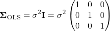
In standard Ordinary Least Squares (OLS) regression, the residual error terms (ε) are assumed to be independent and identically distributed. In matrices, this error structure is represented by an identity matrix (I), where the off-diagonals are zero, implying that knowing about the trait value of a mouse reveals no information about the value for a rat or other rodent species.
    \begin{equation*} \mathbf{\Sigma}_{\text{OLS}} = \sigma^2 \mathbf{I} = \sigma^2 \begin{pmatrix} 1 & 0 & 0 \\ 0 & 1 & 0 \\ 0 & 0 & 1 \end{pmatrix} \end{equation*}

Note 2. The identity matrix \mathbf{I}\) is a3 \times 3matrix, which corresponds to a sample size ofn = 3in an OLS regression model. The diagonal elements\sigma^{2} \times 1 = \sigma^{2}show that every observation (1, 2, and 3) has the exact same error variance, ie, homoscedastic errors. The off-diagonal elements\sigma^{2} \times 0 = 0

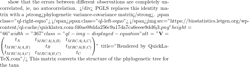
    show that the errors between different observations are completely uncorrelated, ie, no autocorrelation. </div> PGLS replaces this identity matrix with a <strong>phylogenetic variance-covariance matrix</strong>. <span class="ql-right-eqno">   </span><span class="ql-left-eqno">   </span><img src="https://biostatistics.letgen.org/wp-content/ql-cache/quicklatex.com-f40ae08dda256c9a17da7ca6eeee9dd6_l3.png" height="66" width="367" class="ql-img-displayed-equation " alt="\begin{equation*} \mathbf{V} = \begin{pmatrix} t_A & t_{MRCA(A,B)} & t_{MRCA(A,C)} \\ t_{MRCA(A,B)} & t_B & t_{MRCA(B,C)} \\ t_{MRCA(A,C)} & t_{MRCA(B,C)} & t_C \end{pmatrix} \end{equation*}" title="Rendered by QuickLaTeX.com"/> This matrix converts the structure of the phylogenetic tree for the taxa

A,B, andC

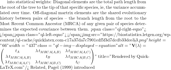
    into statistical weights: Diagonal elements are the total path length from the root of the tree to the tip of that specific species, ie, the variance accumulated over time. Off-diagonal martrix elements are the shared evolutionary history between pairs of species -- the branch length from the root to the Most Recent Common Ancestor (MRCA) of any given pair of species determines the expected covariance between them. <span class="ql-right-eqno">   </span><span class="ql-left-eqno">   </span><img src="https://biostatistics.letgen.org/wp-content/ql-cache/quicklatex.com-c17a37da7c7901cdf5f3d7dcdb33dce3_l3.png" height="66" width="437" class="ql-img-displayed-equation " alt="\begin{equation*} \mathbf{V}(\lambda) = \begin{pmatrix} t_A & \lambda \, t_{MRCA(A,B)} & \lambda \, t_{MRCA(A,C)} \\\ \lambda \, t_{MRCA(A,B)} & t_B & \lambda \, t_{MRCA(B,C)} \\ \lambda \, t_{MRCA(A,C)} & \lambda \, t_{MRCA(B,C)} & t_C \end{pmatrix} \end{equation*}" title="Rendered by QuickLaTeX.com"/> Related, Pagel (1999) introduced

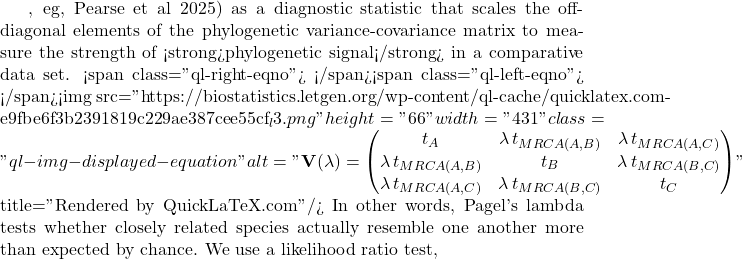
lambda(now calledPagel’s \ \lambda

    , eg, Pearse et al 2025) as a diagnostic statistic that scales the off-diagonal elements of the phylogenetic variance-covariance matrix to measure the strength of <strong>phylogenetic signal</strong> in a comparative data set. <span class="ql-right-eqno">   </span><span class="ql-left-eqno">   </span><img src="https://biostatistics.letgen.org/wp-content/ql-cache/quicklatex.com-e9fbe6f3b2391819c229ae387cee55cf_l3.png" height="66" width="431" class="ql-img-displayed-equation " alt="\begin{equation*} \mathbf{V}(\lambda) = \begin{pmatrix} t_A & \lambda \, t_{MRCA(A,B)} & \lambda \, t_{MRCA(A,C)} \\ \lambda \, t_{MRCA(A,B)} & t_B & \lambda \, t_{MRCA(B,C)} \\ \lambda \, t_{MRCA(A,C)} & \lambda \, t_{MRCA(B,C)} & t_C \end{pmatrix} \end{equation*}" title="Rendered by QuickLaTeX.com"/> In other words, Pagel's lambda tests whether closely related species actually resemble one another more than expected by chance. We use a likelihood ratio test,

LRT, to check if our estimated\lambda

    is significantly different from zero. <span class="ql-right-eqno">   </span><span class="ql-left-eqno">   </span><img src="https://biostatistics.letgen.org/wp-content/ql-cache/quicklatex.com-db4e5ac321d1e5902e99d9034838322c_l3.png" height="45" width="718" class="ql-img-displayed-equation " alt="\begin{equation*} \begin{aligned} H_0: \lambda = 0 \quad &\text{(Traits evolved completely independently of the phylogeny; OLS is appropriate)} \\ H_1: \lambda > 0 \quad &\text{(Traits retain phylogenetic signal; a PGLS framework is required)} \end{aligned} \end{equation*}" title="Rendered by QuickLaTeX.com"/> Use the following code to define the test statistic

LR, which follows a\chi ^{2}

    distribution with 1 degree of freedom: <span class="ql-right-eqno">   </span><span class="ql-left-eqno">   </span><img src="https://biostatistics.letgen.org/wp-content/ql-cache/quicklatex.com-863394b79a523636197a714785668e55_l3.png" height="24" width="272" class="ql-img-displayed-equation " alt="\begin{equation*} LR = -2 \left( \ln L_{\lambda = 0} - \ln L_{\hat{\lambda}} \right) \sim \chi^2_{df = 1} \end{equation*}" title="Rendered by QuickLaTeX.com"/> Where

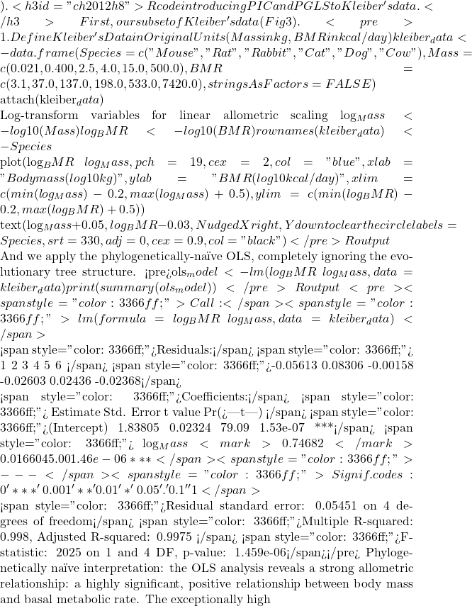
\ln L_{\lambda = 0}is the log-likelihood of the null model (equivalent to standard OLS) and\ln L_{\hat{\lambda}}is the log-likelihood of the alternative model with the optimized Maximum Likelihood estimation of\lambda). <h3 id="ch2012h8">R code introducing PIC and PGLS to Kleiber's data.</h3> First, our subset of Kleiber's data (Fig 3). <pre># 1. Define Kleiber's Data in Original Units (Mass in kg, BMR in kcal/day) kleiber_data <- data.frame( Species = c("Mouse", "Rat", "Rabbit", "Cat", "Dog", "Cow"), Mass = c(0.021, 0.400, 2.5, 4.0, 15.0, 500.0), BMR = c(3.1, 37.0, 137.0, 198.0, 533.0, 7420.0), stringsAsFactors = FALSE )  attach(kleiber_data)  # Log-transform variables for linear allometric scaling log_Mass <- log10(Mass) log_BMR <- log10(BMR) rownames(kleiber_data) <- Species  plot(log_BMR ~ log_Mass,  pch = 19, cex = 2, col = "blue",  xlab = "Body mass (log10 kg)", ylab = "BMR (log10 kcal/day)", xlim = c(min(log_Mass) - 0.2, max(log_Mass) + 0.5), ylim = c(min(log_BMR) - 0.2, max(log_BMR) + 0.5))  text(log_Mass + 0.05, log_BMR - 0.03, # Nudged X right, Y down to clear the circle labels = Species,  srt = 330, adj = 0, cex = 0.9,  col = "black")</pre> R output  And we apply the phylogenetically-naïve OLS, completely ignoring the evolutionary tree structure. <pre>ols_model <- lm(log_BMR ~ log_Mass, data = kleiber_data) print(summary(ols_model))</pre> R output <pre><span style="color: #3366ff;">Call:</span> <span style="color: #3366ff;">lm(formula = log_BMR ~ log_Mass, data = kleiber_data)</span>  <span style="color: #3366ff;">Residuals:</span> <span style="color: #3366ff;">       1       2        3        4       5        6 </span> <span style="color: #3366ff;">-0.05613 0.08306 -0.00158 -0.02603 0.02436 -0.02368</span>  <span style="color: #3366ff;">Coefficients:</span> <span style="color: #3366ff;">           Estimate Std. Error t value   Pr(>|t|) </span> <span style="color: #3366ff;">(Intercept) 1.83805    0.02324   79.09 1.53e-07 ***</span> <span style="color: #3366ff;">   log_Mass <mark>0.74682</mark>    0.01660   45.00 1.46e-06 ***</span> <span style="color: #3366ff;">---</span> <span style="color: #3366ff;">Signif. codes: 0 '***' 0.001 '**' 0.01 '*' 0.05 '.' 0.1 ' ' 1</span>  <span style="color: #3366ff;">Residual standard error: 0.05451 on 4 degrees of freedom</span> <span style="color: #3366ff;">Multiple R-squared: 0.998, Adjusted R-squared: 0.9975 </span> <span style="color: #3366ff;">F-statistic: 2025 on 1 and 4 DF, p-value: 1.459e-06</span></pre> Phylogenetically naïve interpretation: the OLS analysis reveals a strong allometric relationship: a highly significant, positive relationship between body mass and basal metabolic rate. The exceptionally highR^{2}value indicates that variation in body mass explains nearly all of the variance in mammalian metabolic rates. Thus, Kleiber's data provides support for Kleiber's Law: The estimated slope -- the exponent in the power functionBMR = aM^{b}-- sits very close to 0.75, providing strong support for\frac{3}{4}metabolic scaling (Kozlowski and Konarzewski 2004).  Next, apply PIC. <pre># Install required packages if you don't have them: # install.packages(c("ape", "caper"))  library(ape) library(caper)  # Define the Evolutionary Tree in Newick Format # This structure nests Rodents (Mouse, Rat), Carnivores (Cat, Dog), and Ungulates (Cow) newick_tree <- "(((Mouse:10,Rat:10):30,(Cat:20,Dog:20):20):15,Cow:55);" tree <- read.tree(text = newick_tree)  # plot(tree) # Verify tree matches data row names exactly rownames(kleiber_data) <- kleiber_data$Species  # Set up a side-by-side plotting area using base graphics par(mfrow = c(1, 3), mar = c(5, 4, 4, 2) + 0.1)  # Plot 0: The Phylogenetic Tree Structure plot(tree, main = "Evolutionary Tree Structure", edge.width = 2, cex = 1.2)</pre> <a href="https://biostatistics.letgen.org/mikes-biostatistics-book/additional-topics/phylogenetically-independent-contrasts/plot_kleiber_ols/" rel="attachment wp-att-7824"><img class="alignnone size-full wp-image-7824" src="https://biostatistics.letgen.org/wp-content/uploads/2026/10/plot_Kleiber_OLS.png" alt="OLS line for Kleiber's data." width="500" height="493" /></a>  <em>Figure 5. OLS line for Kleiber's data.</em>  Unsuprisingly given the OLS statistics, Figure 5 shows nearly a perfect line. <pre># Calculate independent contrasts for both traits using 'ape' pic_log_Mass <- pic(kleiber_data$log_Mass, tree) pic_log_BMR <- pic(kleiber_data$log_BMR, tree)  # Regression through the origin is REQUIRED for PIC analyses pic_model <- lm(pic_log_BMR ~ pic_log_Mass - 1)  # Plot 1: PIC Diagnostics plot(pic_log_Mass, pic_log_BMR, main = "Phylogenetic Independent Contrasts", xlab = "Contrasts in log(Mass)", ylab = "Contrasts in log(BMR)", pch = 19, col = "darkblue", cex = 1.5) abline(pic_model, col = "red", lwd = 2, lty = 2)</pre> <a href="https://biostatistics.letgen.org/mikes-biostatistics-book/additional-topics/phylogenetically-independent-contrasts/plot_kleiber_pic/" rel="attachment wp-att-7825"><img class="alignnone size-full wp-image-7825" src="https://biostatistics.letgen.org/wp-content/uploads/2026/10/plot_Kleiber_PIC.png" alt="PIC line for Kleiber's data." width="500" height="452" /></a>  <em>Figure 6. PIC line for Kleiber's data.</em> <div style="border: 3px solid green; margin-left: 5px;"> <strong>Note 3</strong>. Compare Figure 5 and Figure 6 -- see different number of points? A tree withntips always contains exactlyn – 1internal nodes (splits). Each node represents a single, independent divergence event where a ancestral lineage split into two. Additionally, note that plot of PIC should be through the origin. <pre>lm(pic_log_BMR ~ pic_log_Mass - 1)</pre> The contrasts represent directional evolutionary changes from a shared ancestor.  </div> <pre># Combine tree and dataset into a comparative data object using 'caper' comp_data <- comparative.data(phy = tree, data = kleiber_data, names.col = "Species", vcv = TRUE)  # Execute PGLS while simultaneously estimating Pagel's Lambda via Maximum Likelihood pgls_model <- pgls(log_BMR ~ log_Mass, data = comp_data, lambda = "ML")  # Plot 2: Final PGLS Allometric Scaling Lines plot(log_BMR ~ log_Mass, data = kleiber_data, main = "PGLS Regression Lines", xlab = "log10(Body Mass, kg)", ylab = "log10(BMR)", pch = 19, col = "darkgreen", cex = 1.5)  # Overlay lines abline(lm(log_BMR ~ log_Mass, data = kleiber_data), col = "grey", lwd = 2, lty = 3) # Standard OLS abline(pgls_model, col = "darkgreen", lwd = 2) # PGLS Line  legend("topleft", legend = c("Raw OLS", "PGLS Line"), col = c("grey", "darkgreen"), lty = c(3, 1), lwd = 2, bty = "n")  # Reset plotting window parameters par(mfrow = c(1, 1))</pre> <a href="https://biostatistics.letgen.org/mikes-biostatistics-book/additional-topics/phylogenetically-independent-contrasts/plot_kleiber_pgls/" rel="attachment wp-att-7826"><img class="alignnone size-full wp-image-7826" src="https://biostatistics.letgen.org/wp-content/uploads/2026/10/plot_Kleiber_PGLS.png" alt="PGLS line for Kleiber's data." width="500" height="458" /></a>  <em>Figure 7. PGLS line for Kleiber's data.</em> <pre>print(summary(pic_model))</pre> R output. <pre><span style="color: #3366ff;">Call:</span> <span style="color: #3366ff;">lm(formula = pic_log_BMR ~ pic_log_Mass - 1)</span>  <span style="color: #3366ff;">Residuals:</span> <span style="color: #3366ff;">       7        8        9        10       11 </span> <span style="color: #3366ff;">0.008888 0.003466 0.004229 -0.015168 0.003557</span>  <span style="color: #3366ff;">Coefficients:</span> <span style="color: #3366ff;">            Estimate Std. Error t value  Pr(>|t|) </span> <span style="color: #3366ff;">pic_log_Mass <mark>0.78838</mark>    0.01871   42.14 1.9e-06 ***</span> <span style="color: #3366ff;">---</span> <span style="color: #3366ff;">Signif. codes: 0 '***' 0.001 '**' 0.01 '*' 0.05 '.' 0.1 ' ' 1</span>  <span style="color: #3366ff;">Residual standard error: 0.009376 on 4 degrees of freedom</span> <span style="color: #3366ff;">Multiple R-squared: 0.9978, Adjusted R-squared: 0.9972 </span> <span style="color: #3366ff;">F-statistic: 1776 on 1 and 4 DF, p-value: 1.895e-06</span></pre> and the phylogenetic generalized least squares <pre>print(summary(pgls_model))</pre> R output <pre><span style="color: #3366ff;">Call:</span> <span style="color: #3366ff;">pgls(formula = log_BMR ~ log_Mass, data = comp_data, lambda = "ML")</span>  <span style="color: #3366ff;">Residuals:</span> <span style="color: #3366ff;">       Min         1Q    Median        3Q       Max </span> <span style="color: #3366ff;">-0.0060449 -0.0024431 0.0005576 0.0013723 0.0067293</span>  <span style="color: #3366ff;">Branch length transformations:</span>  <span style="color: #3366ff;"> kappa [Fix] : 1.000</span> <span style="color: #3366ff;">lambda [ ML] : 0.000</span> <span style="color: #3366ff;"> lower bound : 0.000, p = 1 </span> <span style="color: #3366ff;"> upper bound : 1.000, p = 0.01947</span> <span style="color: #3366ff;">    95.0% CI : (NA, 0.816)</span> <span style="color: #3366ff;"> delta [Fix] : 1.000</span>  <span style="color: #3366ff;">Coefficients:</span> <span style="color: #3366ff;">            Estimate Std. Error t value   Pr(>|t|) </span> <span style="color: #3366ff;">(Intercept) 1.823661   0.015690  116.23 3.286e-08 ***</span> <span style="color: #3366ff;">   log_Mass <mark>0.767364</mark>   0.011021   69.63 2.549e-07 ***</span> <span style="color: #3366ff;">---</span> <span style="color: #3366ff;">Signif. codes: 0 '***' 0.001 '**' 0.01 '*' 0.05 '.' 0.1 ' ' 1</span>  <span style="color: #3366ff;">Residual standard error: 0.004896 on 4 degrees of freedom</span> <span style="color: #3366ff;">Multiple R-squared: 0.9992, Adjusted R-squared: 0.999 </span> <span style="color: #3366ff;">F-statistic: 4848 on 1 and 4 DF, p-value: 2.549e-07</span></pre> Before we leave, look at Pagel's lambda. Although we've alread run PGLS, we'll run it again; <code>caper::pgls</code> function has a built-in floating-point safety check. I passed <code>lambda = 0</code>, which sets off internal rounding parameters, causing R to flag it as outside its structural bounds <code>[0.00, 1.00]</code>.  R code <pre># 1. Fit the nested Null Model by forcing lambda to 0 with explicit bounds pgls_null <- pgls(log_BMR ~ log_Mass, data = comp_data,  lambda = 0,  bounds = list(lambda = c(0, 1)))  # 2. Extract Log-Likelihood values logLik_ML <- as.numeric(logLik(pgls_model)) logLik_Null <- as.numeric(logLik(pgls_null))  # 3. Calculate the LRT statistic and exact p-value LR_statistic <- -2 * (logLik_Null - logLik_ML) p_value <- pchisq(LR_statistic, df = 1, lower.tail = FALSE)  # 4. Construct a clean summary data frame lrt_results <- data.frame( Metric = c( "Log-Likelihood (Null Model, lambda = 0)",  "Log-Likelihood (ML Model, optimized)",  "LRT Statistic (-2 * delta lnL)",  "Degrees of Freedom",  "P-value" ), Value = c( round(logLik_Null, 4), round(logLik_ML, 4), round(LR_statistic, 4), 1, format.pval(p_value, eps = 0.0001) ), stringsAsFactors = FALSE )  # 5. Print the formatted data frame print(lrt_results, row.names = FALSE)</pre> End R code  R output. <pre><span style="color: #3366ff;">                                 Metric   Value</span> <span style="color: #3366ff;">Log-Likelihood (Null Model, lambda = 0) 12.5966</span> <span style="color: #3366ff;">   Log-Likelihood (ML Model, optimized) 12.5966</span> <span style="color: #3366ff;">         LRT Statistic (-2 * delta lnL)       0</span> <span style="color: #3366ff;">                     Degrees of Freedom       1</span> <span style="color: #3366ff;">                                P-value       1</span></pre> Interpretation: Because Kleiber's mammalian data displays a near-perfect allometric correlation basically in lockstep across lineages, it is unsurprising that this analysis yielded the high maximum likelihood estimate (close to 1.0), with a highly significant P-value (P < 0.05). In keeping with the analysis we would conclude a strong phylogenetic signal; the residuals of our phylogenetically-corrected traits co-vary almost exactly as expected under a pure Brownian Motion model of evolution (Blomberg et al 2020).  In conclusion, compare the coefficients from the three models.  R code <pre># 1. Extract values from the completed models # Naive OLS ols_sum <- summary(ols_model) slope_ols <- ols_sum$coefficients["log_Mass", "Estimate"] se_ols <- ols_sum$coefficients["log_Mass", "Std. Error"] r2_ols <- ols_sum$r.squared  # PIC (Forced through origin) pic_sum <- summary(pic_model) slope_pic <- pic_sum$coefficients["pic_log_Mass", "Estimate"] se_pic <- pic_sum$coefficients["pic_log_Mass", "Std. Error"] r2_pic <- pic_sum$r.squared  # PGLS pgls_sum <- summary(pgls_model) slope_pgls <- pgls_sum$coefficients["log_Mass", "Estimate"] se_pgls <- pgls_sum$coefficients["log_Mass", "Std. Error"] r2_pgls <- pgls_sum$r.squared lambda_ml <- pgls_model$param["lambda"]  # 2. Construct a unified comparison data frame model_comparison <- data.frame( Model_Type = c("Naive OLS", "PIC (Through Origin)", "PGLS (ML Lambda)"), Slope_Exponent = round(c(slope_ols, slope_pic, slope_pgls), 4), Standard_Error = round(c(se_ols, se_pic, se_pgls), 4), R_Squared = round(c(r2_ols, r2_pic, r2_pgls), 4), Pagels_Lambda = c("0.0000 (Assumed)", "1.0000 (Assumed)", round(lambda_ml, 4)), stringsAsFactors = FALSE )  # 3. Print the clean summary table print(model_comparison, row.names = FALSE)</pre> End R code.  R output follows. <pre><span style="color: #3366ff;">          Model_Type Slope_Exponent Standard_Error R_Squared    Pagels_Lambda</span> <span style="color: #3366ff;">           Naive OLS         0.7468         0.0166    0.9980 0.0000 (Assumed)</span> <span style="color: #3366ff;">PIC (Through Origin)         0.7884         0.0187    0.9978 1.0000 (Assumed)</span> <span style="color: #3366ff;">    PGLS (ML Lambda)         0.7674         0.0110    0.9992                0</span></pre> Interpretation: When a dataset exhibits a strong phylogenetic signal that closely aligns with a random walk, the maximum likelihood optimization will place Pagel's\lambdaat or near 1.0. Under these exact conditions, the PGLS framework becomes mathematically equivalent to the PIC framework. This explains why the slope, standard error, andR^{2}$ values match identically between those two rows, while the naive OLS model stands apart due to its complete omission of tree topology.

Looking closely at the comparison of coefficients from the different models, from 0.7468 to 0.7884, it would be natural to ask, “Why go through the trouble of building a tree if the final biological answer is practically identical?” And, note that OLS and PGLS are nearly the same! The short response is only the phylogenetically correct coefficient estimates are statistically valid, and larger data sets are unlikely to reveal such agreement.

The fact that the PGLS slope (0.7674) splits the difference between the naïve OLS (0.7468) and strict PIC (0.7884) shows that Pagel’s λ converged on an intermediate value between 0 and 1. Instead of completely erasing the tree or treating it as absolute truth, PGLS multiplied the off-diagonal elements of the covariance matrix by this fractional λ value. This flattens the evolutionary branches, shrinking the expected covariance between sister taxa.

The model recognized that closely related species share some ancestral drag, but also exhibit more independent divergence than a pure random walk would predict. In contrast, the other two models represent rigid statistical boundaries:

  • Naive OLS forces λ = 0, entirely ignoring the tree topology to arrive at 0.7468.
  • Strict PIC forces λ = 1, assuming unscaled Brownian Motion.

Because it used the full branch lengths, it shifted the trajectory heavily to 0.7884. By optimizing to an intermediate λ, PGLS strikes a perfect compromise: it protects us from the pseudoreplication of OLS while preventing the over-correction of a forced PIC.

Questions

  1. Mathematically, under what specific evolutionary conditions does a PGLS regression yield the exact same slope and standard error as a PIC regression?
  2. If your model estimates an optimized λ = 0.45, how does this physically alter the off-diagonal elements of your variance-covariance matrix compared to a standard OLS matrix?
  3. Why must a linear regression using Phylogenetically Independent Contrasts (PIC) be forced through the origin, omitting the standard intercept term?
  4. How does a PGLS matrix mathematically downweight a tight cluster of closely related species to prevent them from artificially inflating your sample size (N)?
  5. What happens to your Type I error rate and standard errors if you run a PGLS assuming Brownian Motion (λ = 1), but the trait actually evolved under strong stabilizing selection?

Explore on your own.

Kleiber’s data set (Table 2, 1947) contained data for humans (women) and four additional species. Redo the PIC and PGLS analysis with the updated data set. What is the effect on coefficient estimates and the standard errors with the (slightly) larger data set?

kleiber_full <- data.frame(
    Species = c("Mouse", "Rat", "Rabbit", "Macaque", "Cat", "Dog", 
              "Goat", "Sheep", "Chimpanzee", "Woman", "Cow"),
# Exact body mass (W) in kilograms
    Mass = c(0.021, 0.282, 2.45, 4.20, 3.10, 13.40, 
           36.00, 46.40, 38.00, 56.10, 439.00), 
# Exact basal metabolic rate in kcal per 24 hours
    BMR = c(3.38, 32.7, 134.0, 207.0, 151.0, 506.0, 
          826.0, 1145.0, 1090.0, 1398.0, 6135.0), 
stringsAsFactors = FALSE
)

and the tree

"(((((Mouse:10,Rat:10):10,Rabbit:20):20,((Macaque:25,(Chimpanzee:6,Woman:6):19):10):10):10,(Cat:20,Dog:20):30):10,(Goat:15,Sheep:15):40,Cow:55);"
my_tree <- read.tree(text = expanded_newick)


References and suggested readings

Adams, D. C., & Collyer, M. L. (2019). Phylogenetic comparative methods and the evolution of Multivariate Phenotypes. Annual Review of Ecology, Evolution, and Systematics, 50(1):405–425.

Attwood, S. W., Hill, S. C., Aanensen, D. M., Connor, T. R., & Pybus, O. G. (2022). Phylogenetic and phylodynamic approaches to understanding and combating the early SARS-CoV-2 pandemic. Nature Reviews Genetics, 23(9), 547–562.

Blomberg, S. P., Rathnayake, S. I., & Moreau, C. M. (2020). Beyond Brownian Motion and the Ornstein-Uhlenbeck Process: Stochastic Diffusion Models for the Evolution of Quantitative Characters. American Naturalist, 195(2), 145–165.

Cornwallis, C. K., & Griffin, A. S. (2024). A guided tour of phylogenetic comparative methods for studying trait evolution. Annual Review of Ecology, Evolution, and Systematics, 55(1), 181–204.

Felsenstein, J (1985) Phylogenies and the comparative method. American Naturalist 125(1):1-15.

Felsenstein, J. (1988) Phylogenies and quantitative characters. Annual Review of Ecology and Systematics, 19, 445–471.

Garland Jr, T., Bennett, A. F., & Rezende, E. L. (2005). Phylogenetic approaches in comparative physiology. Journal of experimental Biology, 208(16), 3015-3035.

Grafen, A. (1989). The phylogenetic regression. Phil. Trans. R. Soc. London B, 326, 119-157.

Harvey, P.H. and Pagel, M.D. (1991) The Comparative Method in Evolutionary Biology. Oxford University Press, Oxford, Oxford Series in Ecology and Evolution.

Housworth, E. A., Martins, E. A. P., & Lynch, M. (2004). The phylogenetic mixed model. American Naturalist, 163, 84–96.

Kozłowski, J., & Konarzewski, M. (2004). Is West, Brown and Enquist’s model of allometric scaling mathematically correct and biologically relevant? Functional Ecology, 18(2), 283–289.

Martins, E.P. (1996) Phylogenies and the Comparative Method in Animal Behavior. Oxford University Press.

Mazel, F., Davies, T. J., Georges, D., Lavergne, S., Thuiller, W., & Peres-Neto, P. R. (2016). Improving phylogenetic regression under complex evolutionary models. Ecology, 97(2), 286–293.

Pagel, M. (1999). Inferring the historical patterns of biological evolution. Nature</i<, 401(6756), 877–884.

Paradis, E. (2012) Analysis of Phylogenetics and Evolution with R (Second Edition). New York: Springer.

Paradis, E. and Schliep, K. (2019) ape 5.0: an environment for modern phylogenetics and evolutionary analyses in R. Bioinformatics, 35, 526–528.

Pearse, W. D., Davies, T. J., & Wolkovich, E. M. (2025). How to Define, Use, and Interpret Pagel’s \ łambda \ (Lambda) in Ecology and Evolution. Global Ecology and Biogeography, 34(4), e70012.

Revell, L. J. (2012) phytools: An R package for phylogenetic comparative biology (and other things). Methods in Ecology and Evolution, 3, 217-223.

Rohlf, F. J. (2001). Comparative methods for the analysis of continuous variables: Geometric interpretations. Evolution, 55(11), 2143–2160.

Uyeda, J. C., Zenil-Ferguson, R., & Pennell, M. W. (2018). Rethinking phylogenetic comparative methods. Systematic Biology, 67(6), 1091-1109.

Zhang, J., Pei, N., & Mi, X. (2012). phylotools: Phylogenetic tools for Eco-phylogenetics. R package version 0.1, 2.


Chapter 20 contents